
Key Takeaways
- Silent Structural Harm: Impacted third molars exert continuous horizontal force against adjacent second molars, causing root resorption, severe decay traps, and localized jawbone deterioration before high-intensity pain occurs.
- Soft Tissue Risks: Partially erupted wisdom teeth create a vulnerable gum flap (operculum) that traps oral debris and bacteria, causing painful infections (pericoronitis), facial swelling, and restricted jaw opening (trismus).
- Sinus & Cyst Complications: Upper wisdom teeth roots can extend into the floor of the maxillary sinus to trigger facial pressure, while lower unerupted teeth can form fluid-filled dentigerous cysts that weaken jaw structure.
- Proactive Evaluation Benefits: Timely diagnostic imaging in early adulthood prevents complex surgical extractions, protects healthy neighboring teeth, and reduces long-term recovery times.
Recognizing early warning signs, such as persistent jaw aching, crowding, swollen gum flaps, and unexpected back-molar decay, is essential to preventing long-term damage to adjacent teeth and jaw structure. When third molars lack adequate room to erupt naturally, evaluating your options for a wisdom tooth extraction in Sarnia allows you to address subterranean structural issues before minor discomfort turns into complex dental emergencies.
Why Should You Consider Wisdom Tooth Extraction in Sarnia Before Severe Pain Starts?
Direct Answer: Proactive wisdom tooth evaluation in Sarnia prevents permanent structural damage to adjacent second molars, stops chronic pericoronitis infections, and minimizes surgical risks by removing impacted third molars before tooth roots fully develop and jawbone density increases.
Wisdom teeth, clinically known as third molars, represent the final set of adult teeth to develop in the human jaw, typically erupting between ages 17 and 25. Evolutionary reductions in human jaw size leave many patients throughout Lambton County without sufficient arch space to accommodate these molars, causing them to remain impacted beneath the gumline or erupt at abnormal angles.
| Impaction Type | Structural Impact on Arch | Primary Clinical Risk |
|---|---|---|
| Horizontal Impaction | Crown grows directly into second molar root | Severe root resorption and second molar loss |
| Angular / Mesial Impaction | Tooth tilts forward against adjacent crown | Deep bacterial pocket and interproximal caries |
| Vertical Impaction | Insufficient vertical room to clear gumline | Recurrent pericoronitis and operculum swelling |
| Distal Impaction | Tooth tilts backward into jawbone ramus | Bone cavitation and potential cyst formation |
When a third molar grows horizontally toward an adjacent second molar, constant mechanical force triggers external root resorption—the progressive destruction of healthy enamel, dentin, and root tissue. Furthermore, partial eruption forms an inaccessible anatomical crevice between the teeth. Bacterial plaque accumulating in this space produces localized acid that causes aggressive, hidden cavities (interproximal caries) on both the third molar and the adjacent second molar.
Clinical evaluation using digital radiography during late adolescence or early adulthood yields superior outcomes because surrounding bone tissue is less dense and tooth roots have not fully anchored near sensory nerve pathways.
What Are the Main Signs Your Wisdom Teeth Are Damaging Surrounding Teeth?
Direct Answer: Primary indicators of second molar damage include sudden front-tooth crowding, persistent food trapping behind back molars, localized gum bleeding, and a deep, throbbing ache during chewing caused by pressure-induced root resorption.
Unchecked physical pressure and bacterial accumulation around third molars frequently cause progressive structural complications across the entire dental arch.
| Symptom | Underlying Pathological Mechanism | Long-Term Structural Risk |
|---|---|---|
| Anterior Crowding & Shifting | Continuous forward pressure along the dental arch | Relapse of orthodontic alignment and bite deviation |
| Food Trapping & Bleeding | Deep interproximal pockets created by partial eruption | Irreversible decay on second molars requiring crowns or root canals |
| Deep Radiating Jaw Ache | Pressure-induced root resorption and periodontal damage | Loss of second molar stability and localized jawbone erosion |
Bite Shifting or Causing Front Tooth Crowding
Forward pressure from impacted wisdom teeth can disrupt dental alignment across the arch. Patients experiencing this issue often report sudden overlapping of lower incisors or noticeable pressure against previous orthodontic retainers.
Getting Unexpected Cavities Between Your Back Molars
Partially erupted wisdom teeth create extremely tight, angular gaps where food debris and oral bacteria accumulate. Since toothbrush bristles cannot access these crevices, deep bacterial plaque forms undisturbed. This causes hidden, aggressive cavities on the posterior wall of healthy second molars, which can go undetected without routine dental X-rays.
Deep Throbbing Pain Near Your Second Molar Mean
A deep, localized ache near the back of the mouth often indicates that an impacted wisdom tooth is pushing directly into the root system of the second molar. This constant force can cause pressure necrosis—the gradual breakdown of healthy tooth structure and surrounding bone tissue—resulting in chronic pain that worsens during chewing.
How Do Soft Tissue and Gum Symptoms Indicate Wisdom Tooth Complications?
Direct Answer: Soft tissue complications manifest as swollen gum flaps (operculum), unpleasant taste from purulent drainage, persistent halitosis, and restricted jaw opening (trismus) resulting from localized bacterial infections known as pericoronitis.
When a third molar only partially breaks through the gum tissue, a flap of soft tissue called an operculum often hangs over the crown. Food debris and oral bacteria easily become trapped beneath this flap, creating an ideal environment for localized bacterial infections known as pericoronitis.
| Clinical Stage | Diagnostic Indicators | Associated Soft Tissue Pathology |
|---|---|---|
| Localized Infection | Gum redness, swelling, purulent exudate | Pericoronitis restricted to operculum flap |
| Muscular Spasm | Painful, restricted mouth opening | Masseter muscle inflammation (trismus) |
| Deep Space Spread | Facial swelling, difficulty swallowing | Submandibular cellulitis requiring urgent care |
When localized gum inflammation from pericoronitis goes untreated, it can spread into surrounding facial tissues and the primary jaw muscle (masseter muscle). This muscular reaction causes trismus, a painful stiffness that severely limits a patient’s ability to open their mouth, making eating and basic oral hygiene difficult.
Can Impacted Wisdom Teeth Cause Sinus Issues or Cysts?
Direct Answer: Yes. Upper wisdom teeth roots grow near the maxillary sinus floor, causing facial pressure and congestion, while lower unerupted wisdom teeth can develop fluid-filled dentigerous cysts that hollow out jawbone tissue.
Impacted wisdom teeth can affect structures beyond the immediate dental arch due to their close proximity to facial anatomy.
| Complication | Anatomical Location | Primary Diagnostic Indicator |
|---|---|---|
| Maxillary Sinus Irritation | Upper third molar roots & sinus floor | Unilateral cheek pressure & nasal congestion |
| Dentigerous Cyst | Lower unerupted tooth crown sac | Radiographic jawbone cavitation & root displacement |
| Jaw Structure Weakening | Mandibular body or ramus | High susceptibility to pathological bone fractures |
How Do Upper Wisdom Teeth Affect Your Sinuses?
The roots of upper third molars develop in close proximity to the floor of the maxillary sinus cavity. As these wisdom teeth push upward against restricted bone space, they can irritate the sinus membrane. Patients frequently mistake this mechanical pressure for chronic sinus infections, experiencing symptoms such as:
- Persistent upper facial pressure directly beneath the cheekbones.
- Single-sided nasal congestion that does not respond to allergy or cold medications.
- Continuous dull aches in the upper back teeth during atmospheric pressure shifts.
What Are Dentigerous Cysts and Jawbone Erosion?
When a wisdom tooth remains completely trapped inside the jawbone, the developmental sac surrounding its crown can fill with fluid, forming a dentigerous cyst. As the cyst slowly expands, it hollows out surrounding jawbone tissue and pushes adjacent tooth roots out of position. Left untreated, a large dentigerous cyst can severely weaken jaw integrity, increasing the risk of spontaneous fractures.
How Do Dentists Diagnose Wisdom Tooth Damage Before It Worsens?
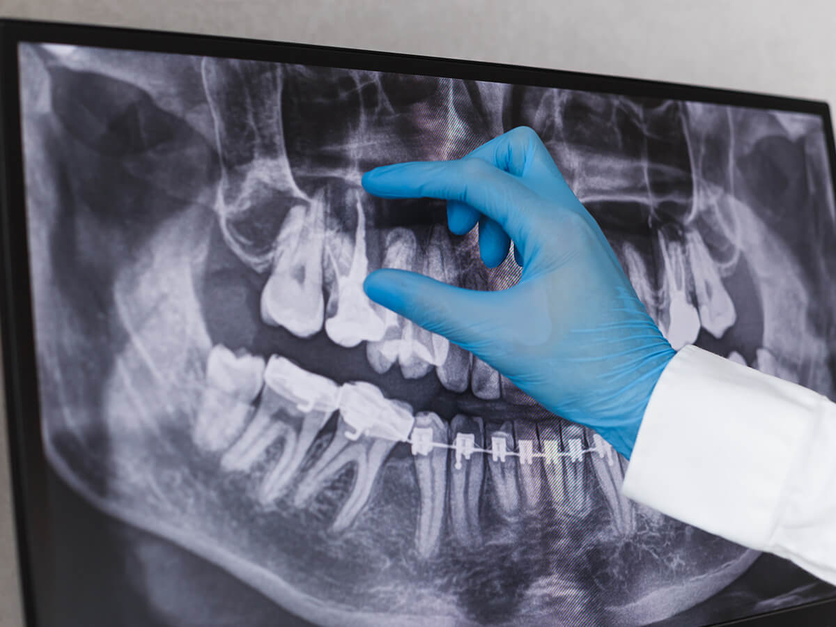
Direct Answer: Dentists diagnose hidden wisdom tooth damage using panoramic X-rays (OPG) for overall arch mapping, 3D Cone Beam Computed Tomography (CBCT) for nerve path location, and periodontal probing to measure deep gum pockets.
Diagnostic tools ensure accurate treatment planning before active symptoms become surgical emergencies.
| Diagnostic Tool | Clinical Purpose | Structural Insight Provided |
|---|---|---|
| Panoramic X-Ray (OPG) | 2D full-arch overview | Tooth impaction angle & neighboring molar health |
| 3D CBCT Imaging | 3D volumetric scanning | Nerve proximity (inferior alveolar nerve) & sinus depth |
| Periodontal Probing | Physical pocket measurement | Bone loss depth between second and third molars |
What to Expect During a Wisdom Tooth Evaluation
A clinical evaluation includes a symptom review, digital diagnostic imaging, a soft-tissue examination, and a personalized discussion of comfortable local anesthesia or oral sedation options tailored to your needs.
| Evaluation Phase | Key Clinical Actions | Patient Expectations |
|---|---|---|
| 1. Symptom Review | Document pain levels, swelling history, and bite shifts | Clear review of current discomfort and health history |
| 2. Diagnostic Imaging | Capture panoramic digital X-rays or 3D CBCT scans | Quick, non-invasive digital imaging process |
| 3. Clinical Examination | Check soft tissue health, operculum status, and decay | Physical assessment of back teeth and jaw mobility |
| 4. Treatment Planning | Outline surgical timeline and review sedation options | Custom care plan with clear recovery guidelines |
Schedule Your Wisdom Tooth Evaluation at Lambton Family Dental
Protecting your long-term oral health starts with identifying wisdom tooth complications before they cause lasting damage to surrounding teeth and jawbone structure. Our team provides clear evaluations and surgical care.
If you are experiencing back molar discomfort, jaw stiffness, or shifting teeth, booking a comprehensive clinical evaluation ensures you receive personalized care in a welcoming environment. Call our family dentist today at 519-344-5747 to schedule your wisdom tooth consultation.
Frequently Asked Questions
1.Why is a single wisdom tooth evaluation rarely sufficient for long-term monitoring?
Wisdom teeth develop over several years, with root structures lengthening and bone density increasing throughout early adulthood. A single X-ray captured during early adolescence cannot predict future eruption paths, making periodic diagnostic evaluations essential to track structural changes over time.
2. What is cumulative dental care, and how does it apply to wisdom tooth management?
Cumulative dental care involves combining proactive diagnostic imaging, ongoing hygiene appointments, and timely surgical interventions to protect long-term oral health. Applying this approach to wisdom teeth prevents minor issues—like partial eruption or mild pericoronitis—from turning into complex dental emergencies that harm neighboring teeth.
3. How long does surgical recovery take following a wisdom tooth extraction?
Initial surface healing and soft tissue recovery typically take between 7 and 14 days. Most patients can resume normal work, school, and light daily activities within 3 to 5 days after surgery. Deeper bone tissue remodeling inside the extraction socket continues naturally under the gumline for several months.